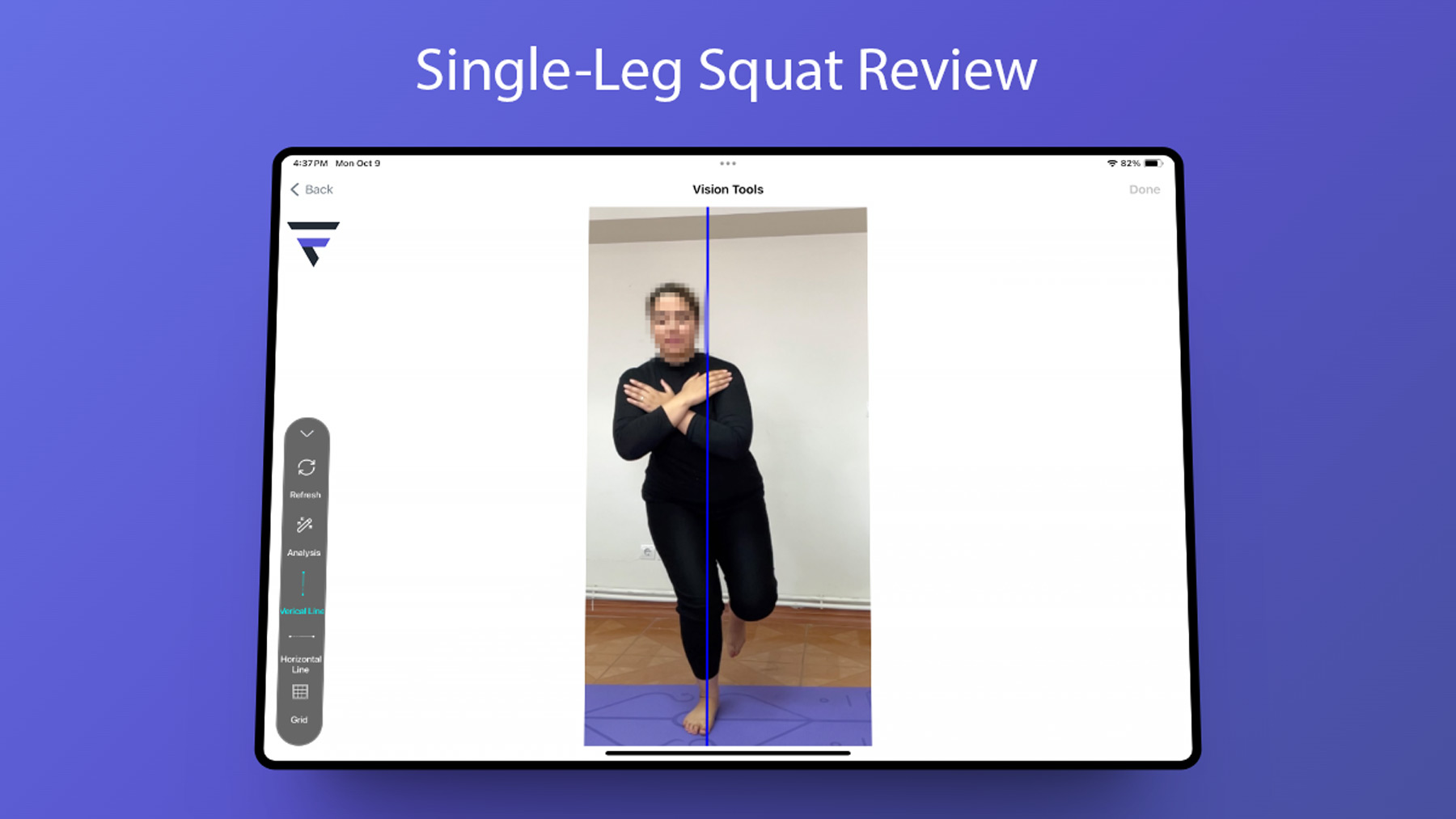
Kinematic review
of
Single-Leg Squat(SLS)
In men and women
The knee joint is primarily designed for flexion and extension movements but also allows minor
abduction, adduction, and lateral sliding. Exceeding normal movement ranges in these directions can be
pathological, potentially leading to skeletal disorders, and thus warrants careful evaluation and
resolution.
The cruciate ligament, vital for knee stability, is also prone to injuries. To prevent such injuries,
it’s
essential to assess the kinematic indicators of the knee, thigh, and ankle. If there’s a risk,
preventative measures should include a regimen to strengthen muscles and improve posture.
Introduction to a Diagnostic Test:
The Single Leg Squat (SLS) test is a crucial assessment for identifying cruciate
ligament injuries and the risk of rupture. During the test, the individual performs a squat on the
injured
leg only, while the uninjured leg remains inactive. The squat should be as deep as possible within the
pain threshold before standing back up. This test helps in evaluating the injured leg’s strength and
stability.
Method
The importance of kinetic results is not hidden from anyone, and the extent of this
importance can be seen in the following review. Below is a balance check with a person tested in the lab
using the FlexiTrace app.
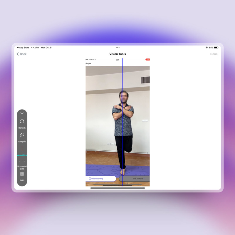

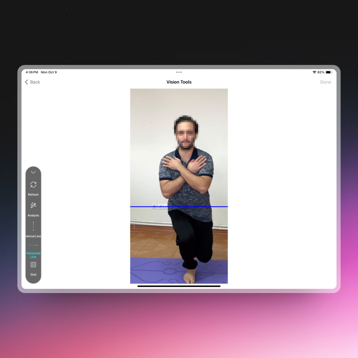
When evaluating pelvic imbalance after a single-leg squat, it’s observed that in an injured individual, the right side of the pelvis is lower than the left side. In contrast, a healthy person’s pelvis remains symmetrical during this exercise. This asymmetry in pelvic alignment during the squat can be an indicator of underlying issues or injuries.
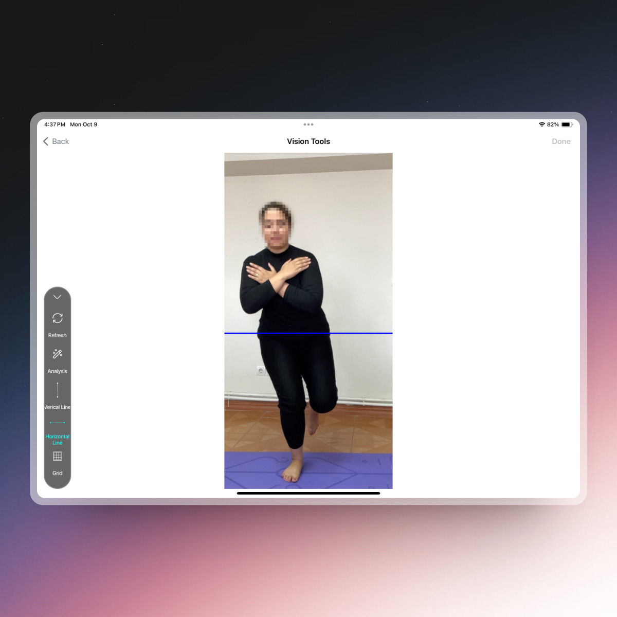
Checking the balance of a healthy person with a knee
injury
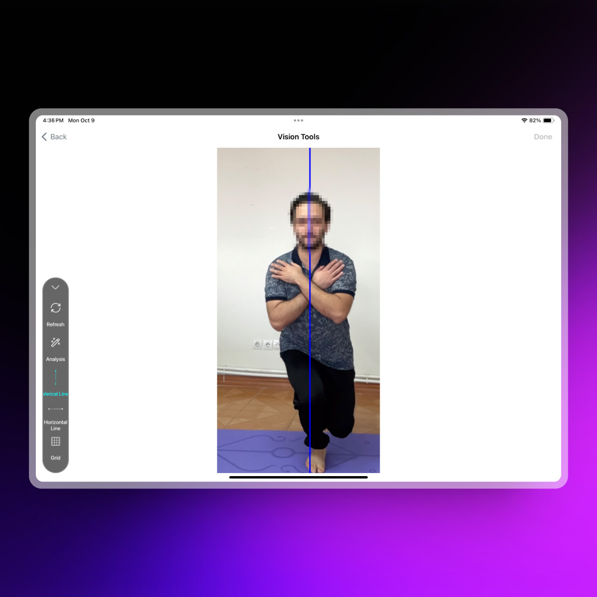
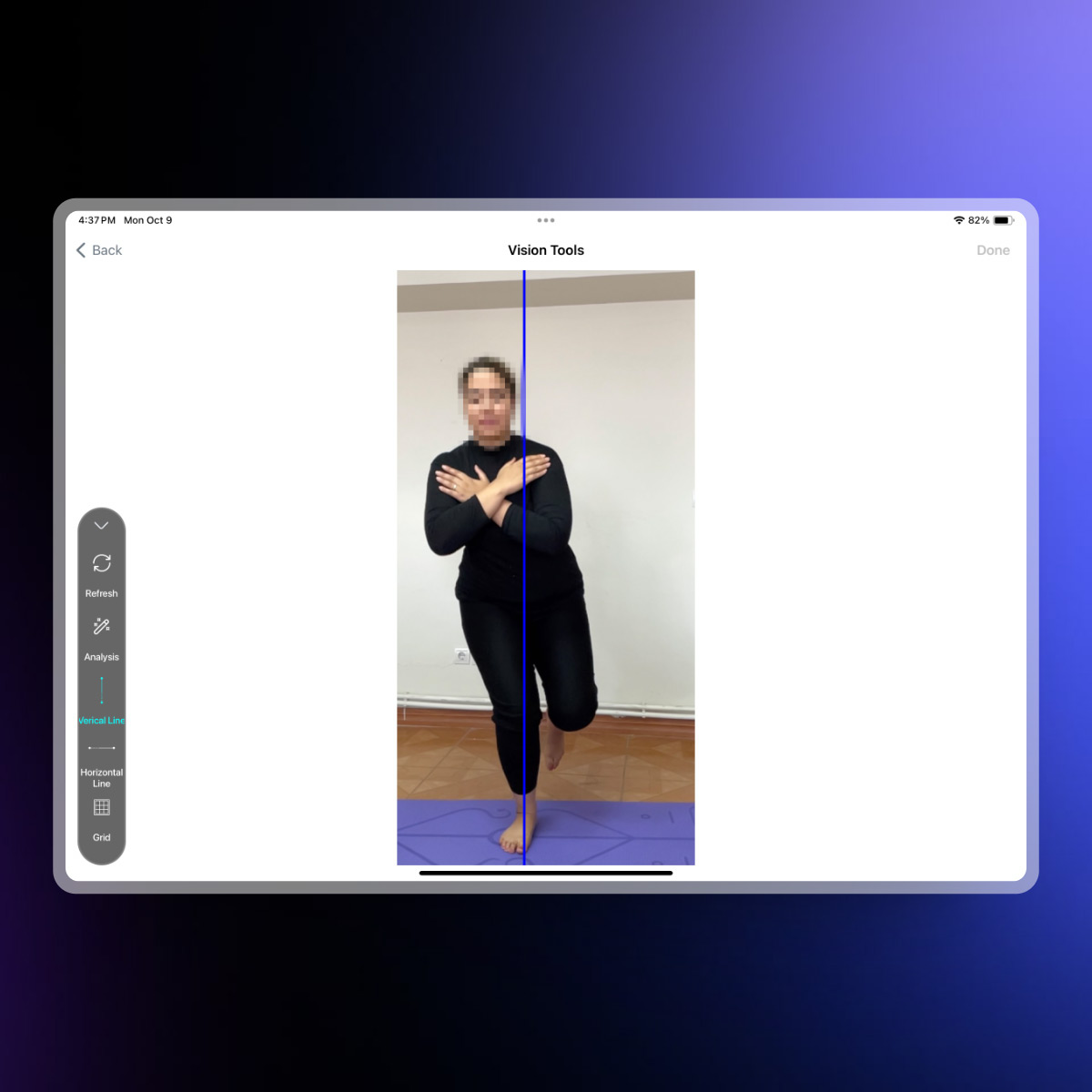
As you can see, a healthy person has kept the balance in the centerline of the body, but a person with a knee injury has deviated from the balance line and tries to maintain balance by moving the body to the right.
As can be seen in the photo, the ankle, knee, and hip joints in a person with a knee injury are not in a vertical alignment.
Discussion:
An examination of the hip and knee joints during a single-leg squat in both healthy and injured
individuals revealed notable differences. Compared to healthy subjects, injured individuals exhibited
a
greater range of motion in the hip’s frontal plane. Moreover, hip joint instability was evident in the
diagrams, particularly visible in the side view during hip flexion, where healthy individuals showed
more pronounced peaks than their injured counterparts.
After the implementation of corrective exercises, tracking and managing the recovery process is
feasible. This can be achieved by utilizing specific assessment tools and making comparisons with the
baseline results. Such an approach allows for a quantifiable evaluation of the rehabilitation progress
and the effectiveness of the corrective exercises applied.
| All Rights Reserved | FlexiTrace Developers LTD